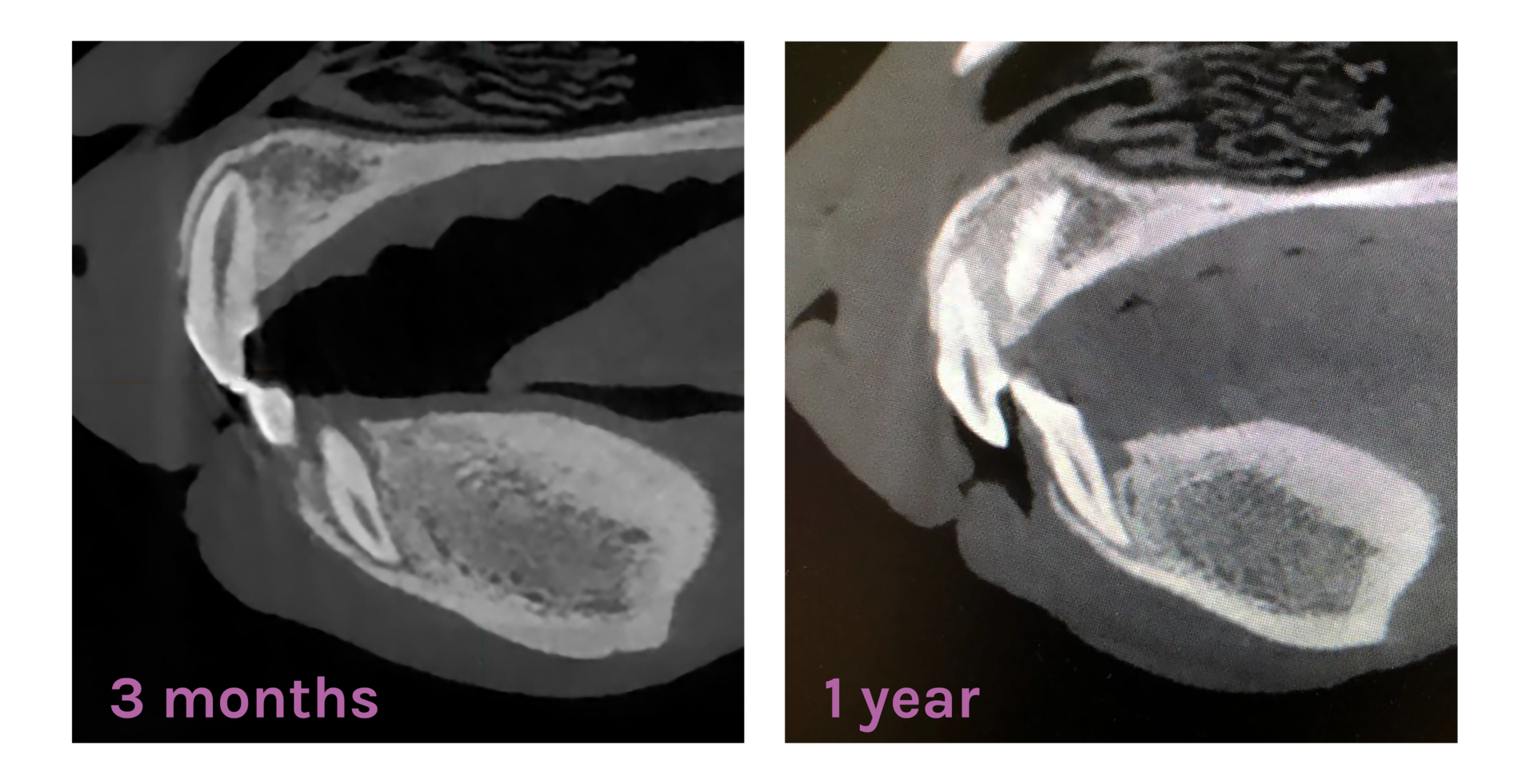
Filling of a miniature Bull Terrier’s canine tooth extraction socket
A lower canine tooth of a 1.5 year old miniature Bull Terrier was removed. The tooth extraction socket was filled with synthetic Adaptos®Vet bone graft granules. The socket was closed with stitches and covered with a biodegradable polymer film as the gingiva was not sufficient to cover the defect. The healing of the defect was uneventful. Three months after the surgery, x-rays showed that the defect had healed and ossified well. Twelve months after the surgery the defect had ossified completely.
Adaptos®Vet for tooth extraction sockets
Particular caution needs to be taken with the tooth extraction of certain teeth: the lower canine tooth (fracture risk in the mandible), lower molar M1 (thin bone wall near the inferior alveolar canal) and upper molar M1 (root of the tooth near the eye). Tooth extraction always creates a hole into the mandible and depending on the breed and the size of the extracted tooth, extraction may lead to weakening of the mandible bone and therefore expose the bone to fractures. For that reason, filling of the socket with a synthetic bone graft that supports ossification of the extraction site, may prevent weakening of mandible bone as well as reduce the risk of fracturing.